understanding the mechanics of nerve repair
There are different ways to repair peripheral nerve gaps, but the size of the bridging material used matters. For example, the sural nerve is a common donor nerve, but it tends to be smaller in diameter,1 often resulting in the need to use multiple strands of the nerve as a graft to match the diameter of the injured nerve. Avance® Nerve Graft is available in a range of diameters, so it can be matched to the diameter of the nerve being repaired. Keep reading to learn more.
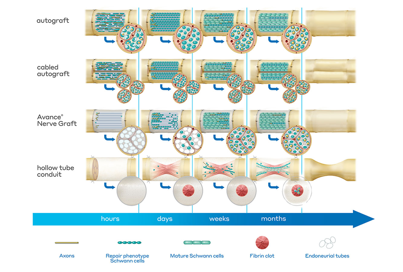
Nerve autograft
- Hours: Autograft tubes are filled with debris and cells from the harvested site; the graft is revascularized and cells begin to clear out tube debris and prepare the structure as a scaffold for regenerating axons.
- Days: Tubes clear, Schwann cells line up and regenerating axons seek tubes to grow; axon regeneration is well distributed through the entire cross section of the graft due to tube organization (scaffolding) in the nerve tissue.
- Weeks: Regenerating axons have found a tube and are close to bridging the entire length of the nerve gap. Over time, these axons regenerate across the entire length of the graft, then continue on to the target site.
- Months: Axons continue to mature and remyelinate, resulting in a new nerve segment that, depending on the injury and the nerve being repaired, may restore nerve function.2
Avance Nerve Graft
- Hours: Axons may begin sprouting; the decellularized matrix begins to revascularize and repopulate with cells.
- Days: Schwann cells infiltrate from both stumps, grow into the empty endoneurial tubes and align, forming structures to help guide axon regeneration across the nerve gap.
- Weeks: Many endoneurial tubes fill with regenerating axons that have remyelinated. Axon regeneration is well distributed throughout the entire cross section of the graft due to the organization of endoneurial tubes throughout the tissue matrix. Over time these axons regenerate across the entire length of the graft and continue to the target site.
- Months: Axons continue to mature and remyelinate, resulting in a new nerve segment that, depending on the injury and nerve being repaired, may restore nerve function.3-6
Hollow tube conduit
- Hours: Fluid containing inflammatory cells and secreted trophic factors fill the conduit.
- Days: A fibrin bridge forms inside the tube; regeneration requires formation of the fibrin matrix, which acts as a physical bridge across the nerve gap; the thickness and quality of the cable is inversely related to the length of the conduit and often does not fill the entire volume of the tube, or does not even form if the gap length is too long.
- Weeks: Schwann cells infiltrate from both stumps and grow along the fibrin cable; some line up and form aligned bands, making a structure that can physically guide regenerating axons across the nerve gap; axon regeneration is centralized to where the fibrin cable forms; some Schwann cell tubes remain empty, and any regenerating axons that do not locate a Schwann cell tube to guide them get pruned back and die. The fibrin bridge breaks down.
- Months: Axons that are able to find and regenerate within an Schwann cell tube grow across the entire length of the conduit and continue on to the target site.3 Depending on the size of the nerve discontinuity and injury, adequate nerve repair can be achieved, but that often results in a thinner nerve segment with variable functionality.7
connect with a nerve rep
There’s only a short form between you and our nerve product team who can help you get more information about our nerve repair solutions.
references
- Ortiguela ME, et al. Anatomy of the sural nerve complex. J Hand Surg Am. Nov 1987; 12(6): 1119-1123.
- Lundborg G. A 25-year perspective of peripheral nerve surgery: evolving neuroscientific concepts and clinical significance. J Hand Surg. 2000;25A:391-414.
- Min Q, et al. Migrating Schwann cells direct axon regeneration within the peripheral nerve bridge. Glia. Feb 2021; 69(2): 235-254.
- Safa B, et al. Peripheral nerve repair throughout the body with processed nerve allografts: Results from a large multicenter study. Microsurgery. Jul 2020; 40(5): 527-537.
- Whitlock EL, et al. Processed allografts and type I collagen conduits for repair of peripheral nerve gaps. Muscle Nerve. 2009 39(6):787-799.
- Evans PJ, et al. The peripheral nerve allograft: a comprehensive review of regeneration and neuroimmunology. Prog Neurobiol. 1994;43:187-223.
- Leversedge FJ, et al. A multicenter matched cohort study of processed nerve allograft and conduit in digital nerve reconstruction. J Hand Surg Am. Dec 2020; 45(12): 1148-1156.